|
FORM IN BRAIN IMAGE TAKESHI MATOZAKI
Name: Matozaki T. Professor, Dr. of Engineering. Address: Department of Electronics & Communication Engineering , Musashi Institute of Technology,1-28-1 Tamazutsumi, Setagaya-Ku,Tokyo 158-8557 Japan. E-mail: tmatozak@musashi-tech.ac.jp Fields of interest: Medical Image Processing, Medical Electronics, Human Perception Technology. Awards: IEEE Fellow Award, 1993; Japan Society of ME&BE Achievement Award, 1977 Abstract: For the simulation
of surgical operations, the extraction of the selected region using MR
images is useful. However, this segmentation requires a high level of skill
and experience from the technicians. We have developed an unique automatic
extraction algorithm for extracting three dimensional brain parenchyma
using MR head images. It is named the "three dimensional gray scale clumsy
painter (CP), moves along the contour of the selected region and extracts
the region surrounded by the contour. This method has advantages compared
with the morphological filtering and the region growing method. Previously,
this method was applied to binary images, but there were some problems
in that the results of the extractions were varied by the value of the
threshold level. We introduced gray level information of images to decide
the threshold, and depend upon the change of image density between the
brain parenchyma and CSF. We decided the threshold level by a map of the
vector of templates, and changed the map according to the change of image
density. As a results, the over extracted ratio was improved by 36%, and
the under extracted ratio was improved by 20%.
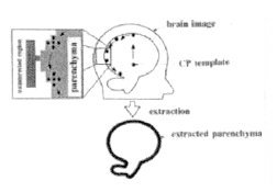
Introduction For the simulation of surgical operations, the extraction of the selected region using MR images is useful. However, this segmentation requires a high level of skill and experience from the physicians. We have developed an unique automatic extraction algorithm for extracting three dimensional brain parenchyma using MR head images. It is named the " three dimensional gray scale clumsy painter method". In this method , a template having the shape of a pseudo-circle, a so called clumsy painter, moves along the contour of the selected region and extracts the region surrounded by the contour. (Fig. 1) .
Method The algorithm of the method is divided into two processes.
The first one's called the edge-searching process and the second one's
the edge -following process. The purpose of the first process is to set
an appropriate initial condition. In the edge-searching process, the initial
point of the template is placed manually so that the template is included
in the interested region. The basis of this method is that the template
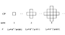
of the approximate circle type (Fig. 2) is made to move
along the contour of region of interest, after the image of the cerebral
parenchyma was transferred to binary image, and that the shape of region
of interest is extracted using the locus.
Fig.1 Schematic diagram of brain parenchyma extraction process
Fig.2 Digital shapes of CP templates in two dimensional CP method
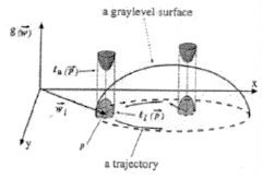
Fig.3 Schematic representation of a
trace of
tl(p)àg(p+wi-1+di)àtu(p) (1) p; element of cp wi-1; cp position after i-1 th moving di; vector of i th movement g(a); image density of position a tl(p); lower map of p tu(p); upper map of p We introduced gray level information of images to decide the threshold ,and it depends upon the change of image density between the brain parenchyma and CSF. We decided the threshold level by the vector of a map of templates, and changed the map according to the change of image density (Fig. 3). However, for the whole image, only one map does be not applied. The map is decided from two thresholds between the lower and the upper level for density. The condition for upper limit is as follows gPD(p+wi-1+di)àtu(p) (2) The condition for lower limit is as follows gT2(p+wi-1+di)àgPD(p+wi-1+di)+K (3)
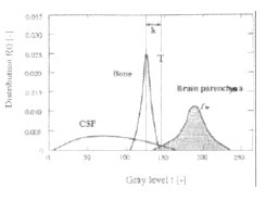
Fig.4 Image density histogram of CSF, back ground, and brain parenchyma
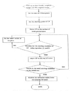
Fig.5 Flow chart of automatic extraction of interested region It is possible to obtain the optimum value of K from the histogram of difference image between the T2 weighted image and Proton density image. In the Fig. 4, the distribution having a peak value at density 127 correspond to the background except brain organ. It is here, and the distribution of cerebral parenchyma is made to be fbp. Mean value of fbp is à, standard deviation à is calculated. Finally, K is decided from this T. The above algorithm is shown in the flow chart. (Fig.
5)
Results & Discussion As a results, the boundary between distribution of the cerebral parenchyma and distribution of the CSF was clarified. It is possible to rightly require the threshold to separating the cerebral parenchyma from the CSF. The proposed technique has introduced the excellent improvement. The extraction accuracy as follows; Excessive extraction ratio was reduced from 1.79% to 1.16%. Lack of extraction ratio was reduced from 2.44% to 1.95%. We shall also display three-dimensional image in order
to visually show the result improving the extraction accuracy.
References Serra, J., " Image Analysis and Mathematical Morphology" London, Academic Press;1982. Serra, J., "Introduction to Mathematical Morphology" Comput, Vision, Graphics, Image Processing 1986;35; pp283-305. Haralik, RM., Sternberg, SR.and Zhuang X.," Image Analysis using Mathematical Morphology" IEEE Trans Pattern Anal. Machine Intell. 1987; pami-9; pp532-550. Sato,M., Ikeguchi T.,
Matozaki,T." An Automatic Algorithm for Removing Uninterested Region in
Image Signals" IEICE Trans on Information and Systems, 1997; E-80-D;
pp63-71.
|